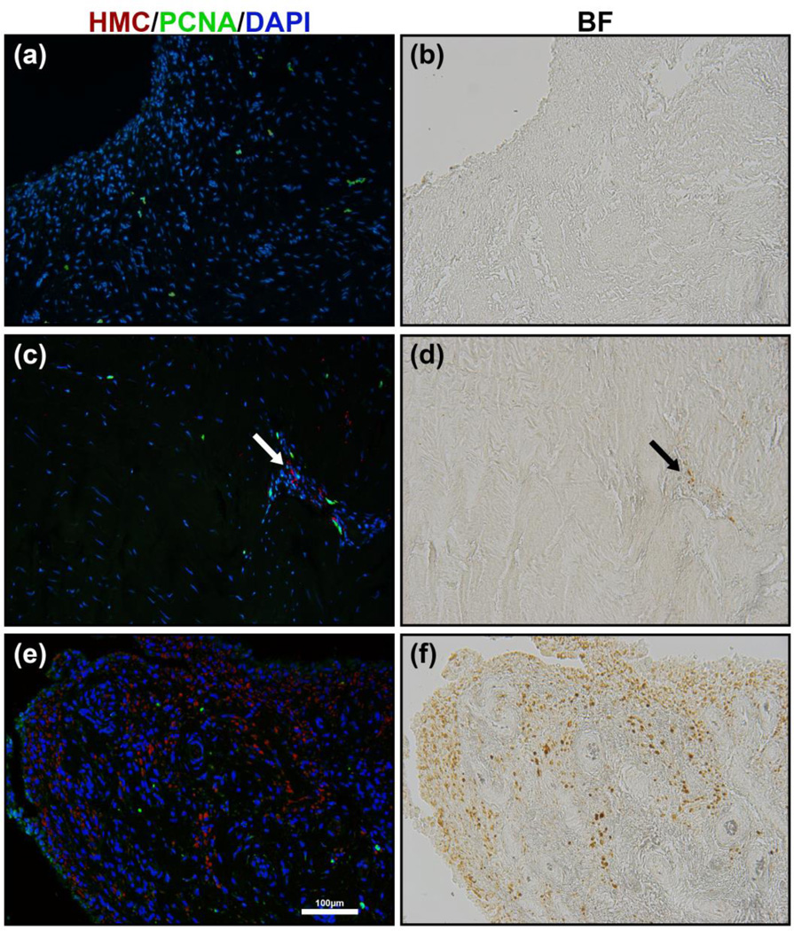
Figure 5.
Proliferating cell nuclear antigen (PCNA) immunostain for cell proliferation of synovial tissue in non-haemophilia patient (a) and haemophilia patient (c & e). Bright-field images (b, d & f) showing the presence of HMCs that correspond to PCNA immunostain (a, c & e) respectively. Proliferating cells were only observed in the fibrous subsurface layer. In haemophilia patients a slight increase in proliferation was observed near haemosiderin-laden macrophage-like cells in the fibrous sub-surface layer (c, arrow). No significant amount of PCNA staining was observed in the surface layer and synovial villi of haemophilia patients (e).

