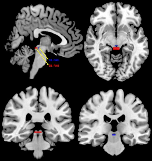
Figure 1.

Dorsolateral and Ventrolateral PAG Regions of Interest. Two box‐shaped masks were created to define both the dorsolateral (DL‐PAG, red; MNI x: 0; y: −32; z: −8.5 plus 6 × 2 × 1.5 mm extensions) and ventrolateral (VL‐PAG, blue; MNI x: 0; y: −27; z: −8 plus 3 × 1 × 1 mm extensions) subdivisions of the PAG. These masks are presented in sagittal (top left), axial (top right), and coronal (bottom) views
