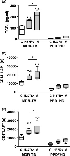
Figure 3.

Enhanced transforming growth factor (TGF)‐β secretion and absolute number of latency‐associated protein (LAP)+ cells in peripheral blood mononuclear cells (PBMC) from multi‐drug‐resistant tuberculosis (MDR‐TB) patients. PBMC from 10 MDR‐TB and six purified protein derivative (PPD)+ healthy donors (HD) were cultured for 48 h alone (control, C) or with Mycobacterium tuberculosis strains and then TGF‐β secretion was determined in PBMC supernatants by enzyme‐linked immunosorbent assay (ELISA) as well as LAP expression was determined in CD14+ and CD4+ cells by FACS. (a). TGF‐β secretion. Results are expressed as pg/ml and medians and 25th–75th percentiles are shown. Statistical differences: *P < 0·05 for control versus M. tuberculosis‐stimulated PBMC or differences among strains (Friedman test followed by Dunn's test); (a) P < 0·05 for M‐ or Ra‐stimulated PBMC from MDR‐TB versus the corresponding data from PPD+ HD (Kruskal–Wallis statistics followed by Dunn's test). (b,c). Number (n) of CD14+ LAP+ cells (b) and CD4+LAP+ cells (c) present in 1× 10⁶ cultured PBMC; medians and 25th–75th percentiles with maximum and minimum values are shown. Statistical differences: *P < 0·05 for treated versus non‐treated PBMC or differences between strains (Friedman test followed by Dunn's test); (a) P < 0·05 for MDR‐TB patients versus PPD+ HD (Kruskal–Wallis statistics followed by Dunn's test).
