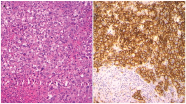
Figure 1.
Histopathologic distinction of SV and NS-HL (a) High power (40×) hematoxylin and eosin stain in SV HL, (b) High power (40×) CD30 immunostain outlining the HRS cells in sheets in SV HL.
HL, Hodgkin lymphoma; HRS, Hodgkin Reed Sternberg; NS-HL, nodular sclerosis Hodgkin lymphoma; SV, syncytial variant.

