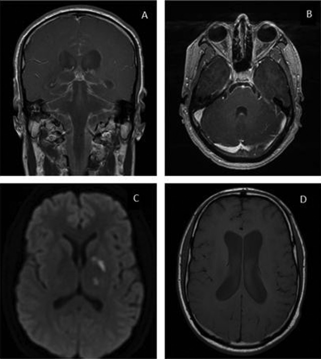
Figure 1.

Magnetic resonance imaging (MRI) of the brain with contrast. Leptomeningeal enhancement was most pronounced in the basilar cisterns (A) and the bilateral fifth and sixth cranial nerves (B) as shown in these gadolinium-enhanced postcontrast T1 images. Foci of reduced diffusion were seen in the left caudothalamic groove, left globus pallidus, and left thalamus (C) as shown in the T2-weighted trace sequence. Moderate communicating hydrocephalus was also present (D) as shown in this T1 sequence.
