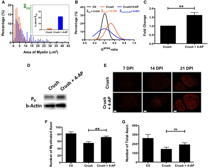
Figure 4. Long‐term/local 4‐AP treatment promoted remyelination and increased the number of myelinated axons.

-
A, BSustained local 4‐AP administration was associated with increased myelin area and close‐to‐normal garea‐ratio compared to untreated group, as determined by analysis of 40 randomly chosen myelinated axons from sections of 4 nerves for each group (P < 0.0001 for saline‐treated vs. 4‐AP treated nerves; ANOVA with post hoc comparisons using two‐tailed unpaired t‐test). 4‐AP‐treated mice had a greater proportion of axons for which the associated myelin area was greater than the mean value for uninjured mice (shown in the green line), with inset figure displaying all axons with values above this mean.
-
C, D4‐AP‐treated nerves also showed increases in the levels of P0 protein as detected by Western blot analysis (**P < 0.01; ANOVA with post hoc comparisons using two‐tailed unpaired t‐test).
-
EIncreases in P0 protein over time also were observed by immunofluorescence analysis at different time points, with P0 protein expression increasing to a greater extent in nerves of mice treated with 4‐AP. Scale bars = 200 μm.
-
F, G4‐AP treatment increased the number of myelinated axons (**P = 0.002; ANOVA with post hoc comparisons using two‐tailed unpaired t‐test; n = 4). Even though the 4‐AP‐treated group also exhibited a greater number of total axons, this difference was not statistically significant. All myelinated and total axons were counted in five randomly chosen grids from each of 4 nerves for each group. This experiment represents a single group of mice of one of three replicates on NCV recovery.
