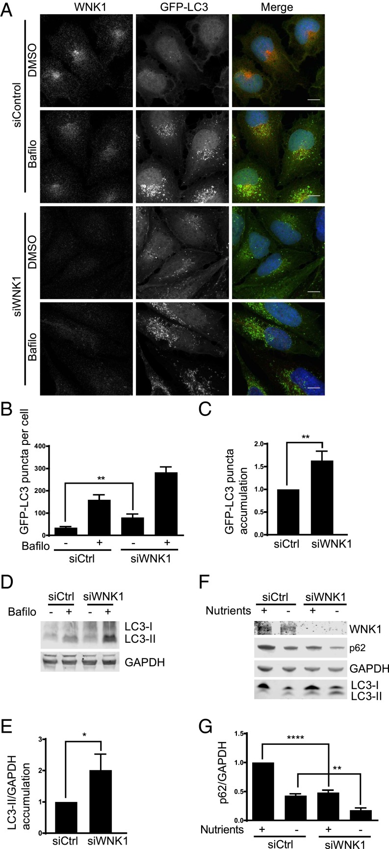
Fig. 1.
WNK1 depletion increases autophagy. (A) U2OS GFP-LC3 cells were treated with 20 nM siControl or siWNK1 oligonucleotides. After 3 d, fresh medium was added containing DMSO or bafilomycin for 4 h. Cells were analyzed by immunofluorescence. The images were deconvoluted, opened in Imaris 8 (Bitplane), and were subjected to background subtraction (filter width, 26.6 μm). The DAPI channel was masked and output was generated for GFP channel to remove the nuclear GFP from the analysis. The puncta-size threshold was set at 0.3 μ and puncta was selected by adjusting the quality in the spots. Then, the puncta were automatically counted by the software. At least 40 cells were selected per condition in each experiment. (Scale bars, 12 μm.) (B) Quantitation of change in number of GFP-LC3 puncta per cell in A. (C) Quantitation of relative change in GFP-LC3 puncta in bafilomycin-treated cells in A. (D) WNK1 was knocked down in U2OS GFP-LC3 cells which were then treated with bafilomycin as in A. Immunoblots are shown. (E) Quantitation of relative change of LC3-II in bafilomycin-treated cells in D. (F) HeLa cells were treated with 20 nM siControl or siWNK1#2 for either 3 d or repeated after 2 d. Before lysis cells were incubated in medium without or with nutrients for 4 h. Proteins were analyzed by immunoblotting. (G) Quantitation of relative change of p62 in F. *P < 0.05, **P < 0.01, ****P < 0.0001.

