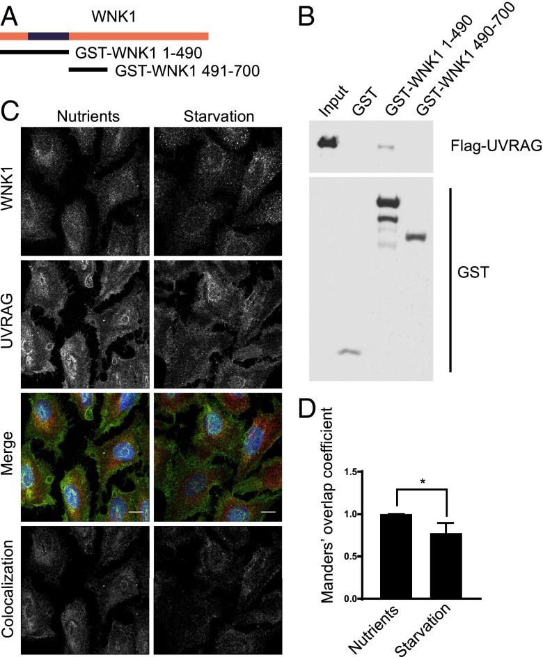
Fig. 4.
WNK1 colocalizes and interacts with UVRAG. (A) Rat WNK1 fragments. WNK1 1–490 encompasses the kinase domain, residues 193–490. (B) Recombinant Flag-UVRAG proteins were captured in vitro with the indicated GST proteins. Binding was detected by immunoblotting and chemiluminescence as in Fig. 3E. (C) U2OS cells were plated on glass coverslips. After 3 d, fresh normal or starvation medium was added for 4 h. Cells were analyzed by immunofluorescence. Images were deconvoluted and corrected for background as in Fig. 2. Colocalization thresholds for WNK1 and UVRAG channels were calculated and a colocalization channel was generated by the Imaris 8 software. The colocalization image shown was captured by the software. (Scale bars, 12 μm.) (D) Quantitation of the change of colocalization between WNK1 and UVRAG in C, using the relative Manders’ coefficient. *P < 0.05.

