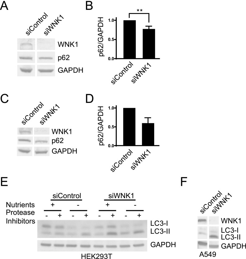
Fig. S1.
WNK1 depletion increases autophagy in mutliple cell lines. (A) HeLa cells were treated with 20 nM siControl or siWNK1 #3 oligonucleotides. After 3 d, the medium was replaced with fresh medium for 4 h. Cells were then lysed and proteins analyzed by immunoblotting. (B) Quantitation of the relative change of p62 in A. **P < 0.01. (C) HeLa cells were treated with 20 nM siControl or siWNK1 #4 oligonucleotides. After 3 d, the medium was replaced with fresh medium for 4 h. Cells were lysed and proteins analyzed by immunoblotting. n = 2. (D) Quantitation of the relative change of p62 in C. (E) HEK293T cells were treated with 10 nM siControl or siWNK1 #2 oligonucleotides. After 3 d, cells were treated with E-64d/pepstatin A or DMSO for 4 h. Cells were then lysed in (150 mM NaCl, 10 mM Tris pH 7.4, 2 mM EDTA, 1 mM EGTA, 1% Triton X-100, and 0.5% Nonidet P-40 with 1% protease inhibitors), mixed with Laemmli sample buffer, and proteins were analyzed by immunoblotting. Secondary antibodies conjugated with horseradish peroxidase were used for blotting by enhanced chemiluminescence. Other WNK1 knockdown experiments in this cell line yielded similar results. (F) A549 cells were treated with 10 nM siControl or siWNK1#2 oligonucleotides. After 3 d, cells were lysed and proteins analyzed by immunoblotting as in E.

