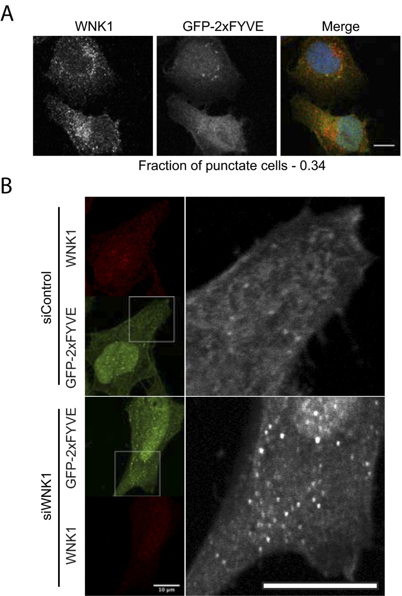
Fig. S2.
WNK1 depletion increases the activity of the PI3KC3 complex. (A) The experiment was performed as described in Fig. 2A in U2OS cells. Cells treated with wortmannin (150 nM) in starvation medium are shown in this figure. (Scale bar, 12 μm.) (B) WNK1 was knocked down by 20 nM siRNA from HeLa cells. The next day, the cells were transfected with a plasmid expressing a GFP-2xFYVE domain. After 2 more days, the medium was replaced by fresh medium for 4 h. Then the cells were analyzed by immunofluorescence on a Zeiss LSM 780. Images shown on the right are enlargements of boxed portions of images on the left. Images shown here are taken from the midplane of the Z stack. (Scale bars, 10 μm.)

