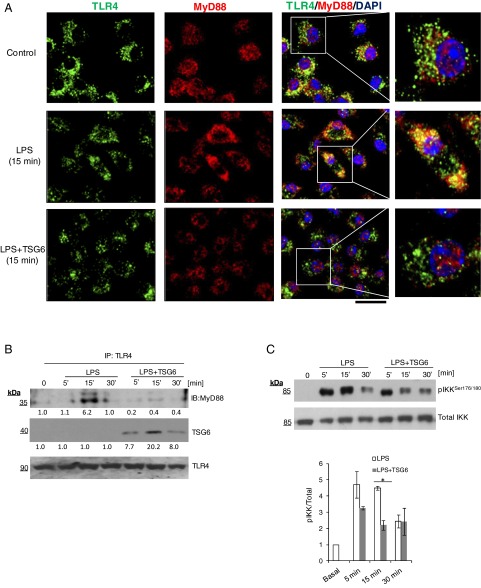
Fig. 2.
TSG6 inhibits the interaction of TLR4 with MyD88 and prevents NF-κB activation. (A) Macrophages cultured from bone marrow of WT mice were challenged with LPS (1 µg/mL) alone or TSG6 (0.1 µg/mL) plus LPS (1 µg/mL) together for 0 and 15 min. Cells were fixed, permeabilized, and stained, and images were acquired by confocal microscopy. Green, TLR4; red, MyD88; blue, DAPI. Strong colocalization of TLR4 with MyD88 was seen after LPS challenge, and this response was prevented by rTSG6. (Scale bar: 20 μm.) (B) WT macrophages cultured from bone marrow were treated with LPS (1 µg/mL) alone or with rTSG6 (0.1 µg/mL) plus LPS (1 µg/mL) for different times. They were then lysed and immunoprecipitated with anti-TLR4 polyclonal antibody (pAb). Immunoprecipitated samples were blotted with anti-MyD88 or anti-TSG6 pAb, and blots were reprobed with anti-TLR4 pAb. A strong interaction of MyD88 with TLR4 was observed at 15 min after LPS treatment, which was abolished by rTSG6. (C) The cell lysates were immunoblotted with anti–phos-Ser177/181-IKKβ pAb. Quantified results are shown as ratio of phosphorylated to total protein (Bottom). rTSG6 treatment decreased LPS-induced phosphorylation of IKKβ. *P < 0.05, LPS vs. LPS + TSG6.

