Fig. 5.
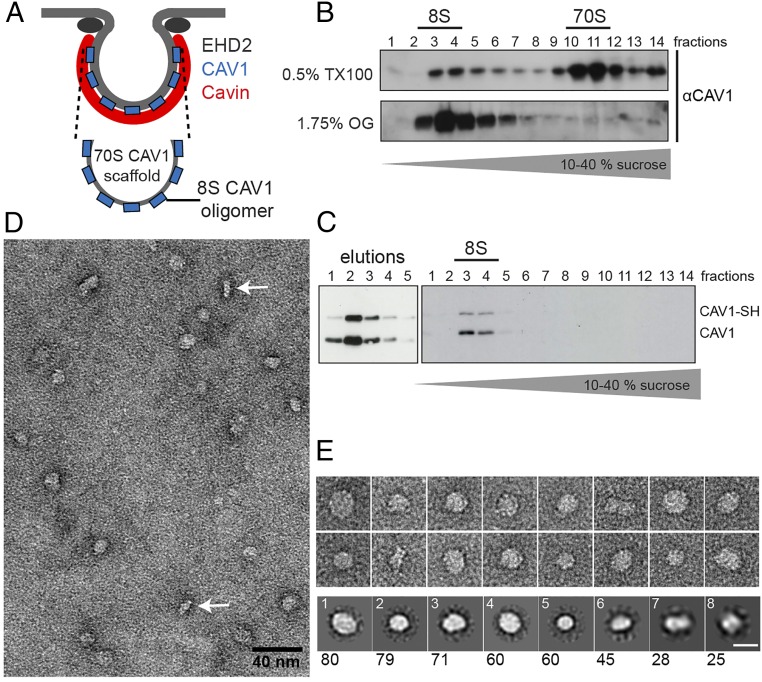
CAV1 8S oligomers are round protein discs. (A) Schematic of the CAV1 70S and 8S complex. (B) Cellular extracts were prepared using 0.5% TX100 or 1.75% octyl-glucoside (OG) and were run through 10–40% sucrose velocity gradients. CAV1 is present in 8S and 70S complexes in TX100 extracts. All CAV1 sediments as 8S complexes in OG extracts. (C) Purification of Strep-HA-CAV1 expressed in HEK293 cells. (Right) SH-CAV1 is coeluted and forms complexes with endogenous CAV1 (eluted fractions 1–5). (Left) Fractions of 10–40% sucrose velocity gradient of purified CAV1, analyzed by SDS/PAGE/Western blot. All CAV1 is present in the 8S oligomer. (D and E) Negative-stain TEM of purified CAV1. (D) Micrograph of CAV1 particles in negative stain. Side views are indicated by white arrows. (E, Upper) Gallery of particles from TEM images. (Lower) 2D class averages from 450 particles, numbered according to the abundance of particles in each class (given below). The majority of CAV1 8S complexes display a circular shape ∼15 nm in diameter. (Scale bar in E: 15 nm.)

