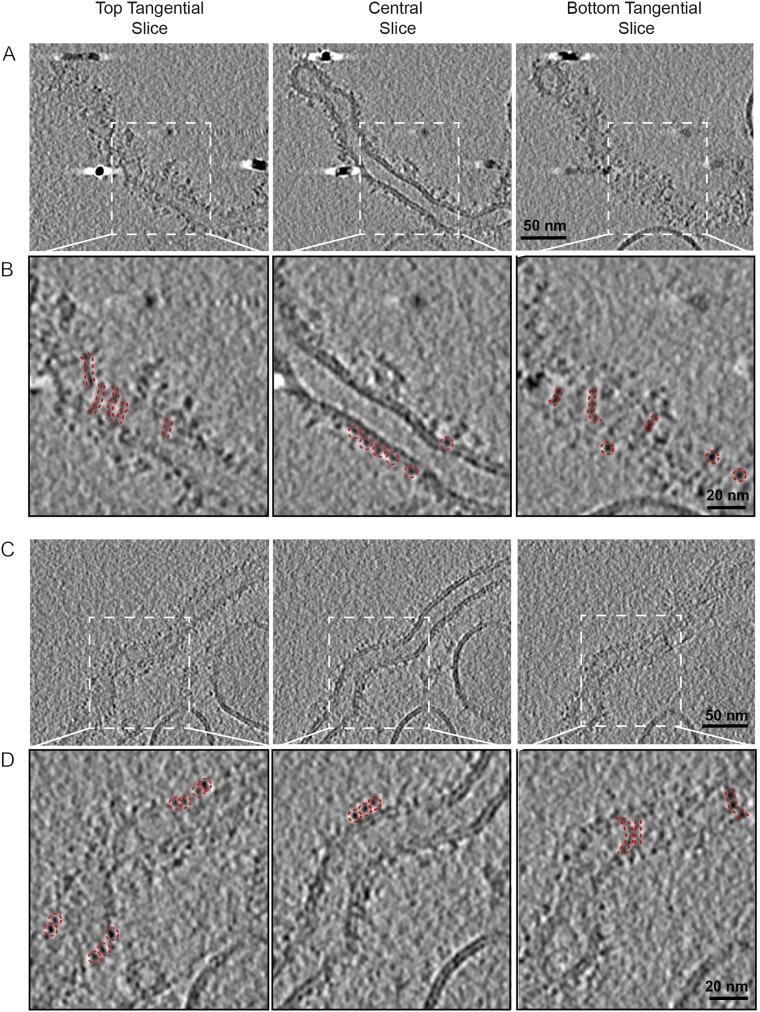
Fig. S4.
Structural characterization by cryoET of Cavin1-Δcc2 liposome binding. Liposomes were incubated with Cavin1-Δcc2 for 60 min and were imaged by cryoET. Panels A and B and panels C and D show two representative events of Cavin1-Δcc2 liposome tubulation. The top, central, and bottom sections of each tomogram are shown. Zoomed views of the boxed areas in the upper rows are displayed in the lower rows. The presence of filaments is seen on the top and bottom tangential views (red dashed lines). The central sections show the cavin coat tightly associated with the membrane (red dashed circle), with individual average densities of 3–4 nm. Liposome composition was 25% DOPS, 15% DOPC, 15% DOPE, and 45% cholesterol. Images were recorded at 300 keV with final calibrated pixel size of 5 Å at defoci of −3.5 to −5 µm.

