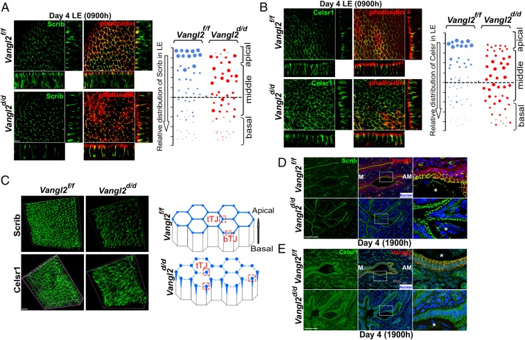
Fig. 2.
Aberrant distribution of PCP proteins in Vangl2d/d luminal epithelia. (A, Left) 3D reconstruction of Scrib localization by IF in ex vivo explants of Vangl2f/f and Vangl2d/d uterine LE on day 4 showing that colocalization of Scrib and phalloidin was lost in Vangl2d/d LE cells. (Scale bar, 10 μm.) (Right) Relative apicobasal distribution of Scrib in Vangl2f/f and Vangl2d/d LE. (B, Left) 3D reconstruction of Celsr1 localization on the morning of day 4. (Scale bar, 10 μm.) (Right) Relative apicobasal distribution of Celsr1 in Vangl2f/f and Vangl2d/d LE. (C, Left) Merged 3D images of IF of Scrib and Celsr1 at the apical surface of Vangl2f/f and Vangl2d/d LE cells on the morning of day 4. (Right) Changes in Scrib and Celsr1 accumulation at the apical surface of Vangl2f/f and Vangl2d/d LE cells. Note that in floxed LE, Scrib and Celsr1 localize to bicellular (bTJ) and tricellular (tTJ) tight junctions, but they are assembled primarily in tricellular junctions in Vangl2d/d LE. (Scale bar, 10 μm.) (D) IF of Scrib in the longitudinal uterine sections of Vangl2f/f and Vangl2d/d mice on the evening of day 4. (Scale bar, 100 µm.) (E) IF of Celsr1 in the longitudinal uterine sections of Vangl2f/f and Vangl2d/d mice on the evening of day 4. (Scale bar, 100 µm.) Asterisks in D and E indicate the location of embryos.

