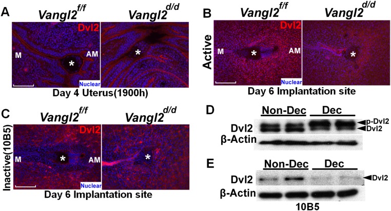
Fig. S7.
Dvl2 localization in puncta in the decidua. (A) IF of Dvl2 at the site of attachment in the evening of day 4. (Scale bar, 100 μm.) (B) IF of Dvl2 in the decidua on day 6. IF results show puncta formation in the primary decidual zone in Vangl2f/f mice but not in Vangl2d/d females. Asterisks indicate the location of embryos. (Scale bar, 200 μm.) (C) IF of nonphosphorylated Dvl2 antibody (10B5) in the decidua on day 6 showing the absence of puncta in both of Vangl2f/f and Vangl2d/d females. (Scale bar, 200 μm.) Asterisks indicate the location of embryos. (D) Phosphorylated Dvl2 levels increased in stromal cells after the induction of decidualization in culture. (E) Nonphosphorylated Dvl2 levels decreased in stromal cells decidualized in vitro.

