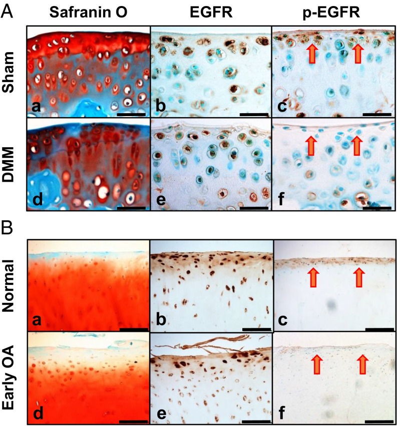
Fig. 1.
EGFR signaling in healthy and diseased articular cartilage. (A) Safranin O staining (a and d) and immunohistochemistry of EGFR (b and e) and p-EGFR (c and f) in tibial articular cartilage of WT mice at 1 mo postsham (a–c) or post-DMM (d–f) surgery. (Scale bars, 50 μm.) (B) Safranin O staining (a and d) and immunohistochemistry of EGFR (b and e) and p-EGFR (c and f) in human joints with healthy (a–c) and early OA (d–f) cartilage. (Scale bars, 200 μm.) Red arrows point to superficial cells.

