Fig. 2.
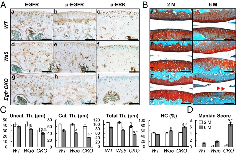
EGFR signaling is important for articular cartilage structure. (A) Immunostaining of EGFR (a, d, and g), p-EGFR (b, e, and h), and p-ERK (c, f, and i) in tibial articular cartilage of 2-mo-old WT (a–c), Wa5 (d–f), and Egfr CKO (g–i) mice. (Scale bars, 100 μm.) (B) Safranin O staining of mouse joints at 2 and 6 mo of age. Red arrows point to reduced cartilage thickness and staining intensity in CKO mice. F, femur; T, tibia. (Scale bars, 100 μm.) (C) Average thicknesses of uncalcified (Uncal. Th.), calcified (Cal. Th.), and total (Total. Th.) articular cartilage in femoral condyle were quantified. The percentage of hypertrophic chondrocyte (HC) in both femoral condyle and tibial plateau was quantified together. (D) Mankin score confirms that CKO mice develop early OA symptoms at 6 mo of age. n = 6 per age per genotype. *P < 0.05, $P < 0.01, &P < 0.001 vs. WT; %P < 0.01, ^P < 0.001 vs. Wa5.

