Fig. 4.
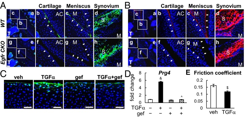
EGFR signaling promotes lubricant secretion from cartilage and meniscus surfaces. (A) Immunofluorescence staining of Prg4 (green) in 1-mo-old WT and CKO joints. The articular cartilage surface (b and f) and meniscus surface (c and g), pointed by white arrows, are magnified images from a and e. Synovium is shown in d and h. AC, articular cartilage; M, meniscus; S, synovium; T, tibia. (Scale bars, 70 μm.) (B) Detection of HA (red) in 1-mo-old WT and CKO joints. The articular cartilage surface (b and f) and meniscus surface (c and g) are magnified images from a and e. Synovium is shown in d and h. (Scale bars, 70 μm.) (C) Immunofluorescence shows that TGFα increases the Prg4 amount (green) at the bovine cartilage explant surface in an EGFR-dependent manner. Blue, DAPI. (Scale bars, 50 μm.) (D) TGFα increased Prg4 mRNA in the top 1-mm region of cartilage explant in an EGFR-dependent manner as assayed by real-time RT-PCR. &P < 0.001 vs. vehicle; ^P < 0.001 vs. TGFα. (E) TGFα treatment reduced the friction coefficient of the articular surface in cartilage explants. n = 5 per group. $P < 0.01 vs. vehicle.

