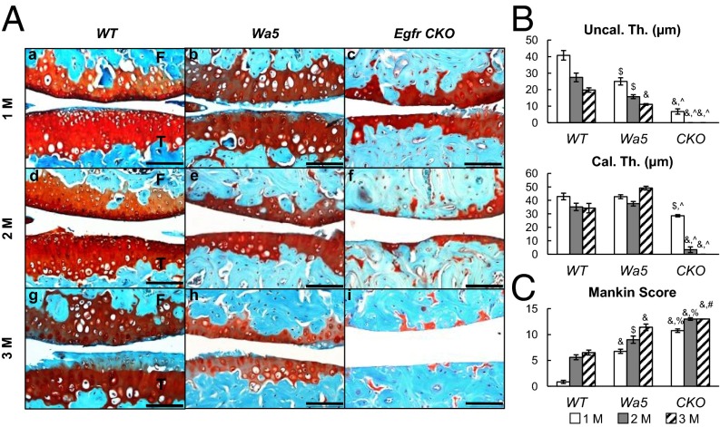
Fig. 6.
EGFR chondrogenic deficiency causes severe OA after DMM surgery. (A) Safranin O staining of WT (a, d, and g), Wa5 (b, e, and h), and CKO (c, f, and i) mouse joints at the medial site at 1 (a–c), 2 (d–f), and 3 (g–i) mo postsurgery. (Scale bars, 100 μm.) (B) Average thicknesses of uncalcified (Uncal. Th.) and calcified (Cal. Th.) cartilage were quantified. n = 6 per age per genotype. $P < 0.01, &P < 0.001 vs. WT; ^P < 0.001 vs. Wa5. (C) The OA severity was measured by Mankin score. $P < 0.01, &P < 0.001 vs. WT; #P < 0.05, %P < 0.01 vs. Wa5. n = 6 per age per group.

