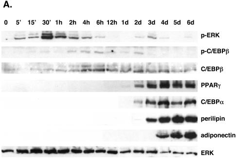
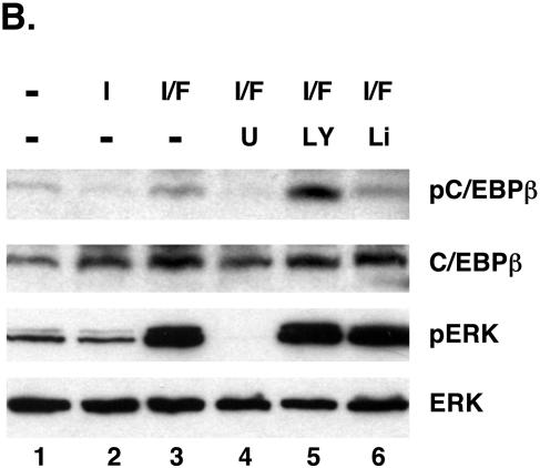
FIG. 1.
Phosphorylation of C/EBPβ at a consensus ERK/GSK3 site during the differentiation of 3T3-L1 preadipocytes. (A) Proliferating 3T3-L1 preadipocytes were cultured in growth medium until they reached confluence. At 2 days postconfluence (day 0), the quiescent cells were exposed to DEX, MIX, FBS, and insulin, and total cellular protein was harvested at the indicated times. Equal amounts of protein from each sample were subjected to Western blot analysis using antibodies specific for phospho-ERK1/2 (p-ERK), phospho-C/EBPβ (p-C/EBPβ), ERK, C/EBPβ, PPARγ, C/EBPα, perilipin, and adiponectin. (B) Confluent 3T3-L1 preadipocytes were exposed to various combinations of 1.67 μM insulin (I), 1 nM FGF-2 (F), 20 μM U0126 (U), 50 μM LY294002 (LY), and 10 mM LiCl (Li) in the presence of MIX and DEX for 4 h, and total cellular protein was subjected to Western blot analysis using antibodies specific for phospho-C/EBPβ (pC/EBPβ), C/EBPβ, phospho-ERK1/2 (pERK), and ERK.


