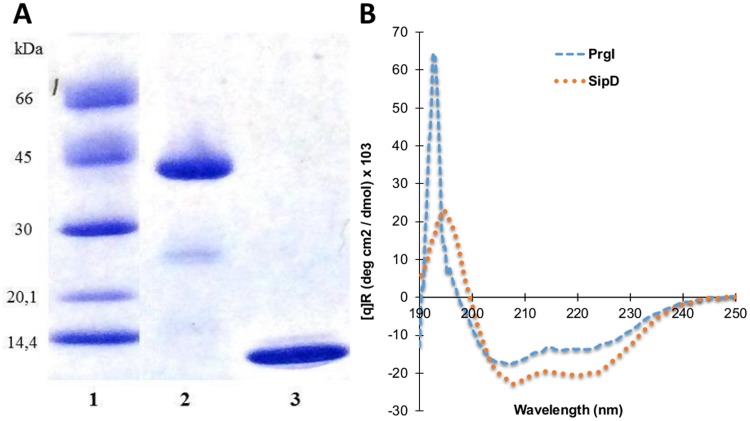
Fig 1. Analysis of recombinant PrgI and SipD proteins.
(A) SDS-PAGE / Coomassie blue staining (reducing conditions) of purified recombinant proteins. polyHis-SipD (38.2 kDa, lane 2) and polyHis-PrgI (9.9 kDa, lane 3) are shown with molecular mass markers in kilodaltons (kDa) (lane 1). (B) Far-UV Circular-Dichroism spectroscopy of recombinant proteins were recorded at 20°C in phosphate buffer at pH 7.4.

