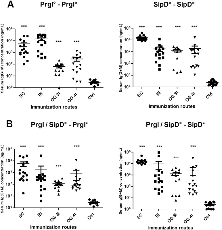
Fig 2. Serum Ig(G+M) concentrations of mice immunized with PrgI or SipD (A) and PrgI/SipD (B).
Serum Ig(G+M) antibodies specific for PrgI (left) and SipD (right) were quantified by sandwich ELISA 2 weeks after the last immunization as described in Materials and Methods. Data represent mean concentrations (ng/mL) and the standard errors (SEM) from 14–16 individual mice per group. Asterisks *** indicate P value< 0.001, comparing the antibody responses using different routes versus control mice. No cross-reactions were observed between PrgI and SipD (data not shown). [°: indicates injected immunogen; *: indicates biotinylated recombinant protein].

