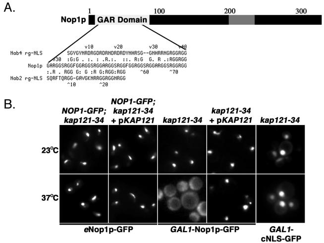
FIG. 2.
Nop1p mislocalizes in kap121-34 cells. (A) Schematic diagram of Nop1p. The white segment highlights the RG-rich GAR domain of Nop1p. The gray segment represents a basic stretch of amino acids (199 to 219; SHRPGRELISMAKKRPNIIP) that is most similar to the previously identified Kap121p NLS sequences. The NLS sequences of Nab2p and Nab4p (27) were compared to full-length Nop1p by using MegAlign (Lipman-Pearson: ktuple, 2; gap penalty, 4; gap length penalty, 12). (B) The distribution of endogenously tagged NOP1 (eNop1p-GFP) and galactose-inducible NOP1GFP (GAL1-Nop1p-GFP) were monitored by direct fluorescence microscopy in kap121-34 cells. NOP1 GFP; kap121-34 cells were visualized after growth to mid-logarithmic phase at the permissive temperature (23°C) and after 3 h of growth at the restrictive temperature (37°C). Although the galactose-inducible NOP1GFP chimera was expressed in kap121-34 cells at 23 or 37°C, eNop1p-GFP remains nucleolar in hap121-34 cells after 3h of growth at 37°C. However, GAL1-Nop1p-GFP accumulates in the cytoplasm of kap121-34 cells when expressed at the nonpermissive temperature. This contrasts with control kap121-34 cells containing a wild-type copy of KAP121 (kap121-34+pKAP121) or kap121-34 cells expressing a GAL1-cNLS-GFP chimera.

