FIG. 4.
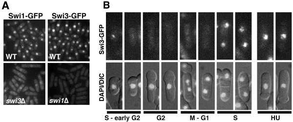
Recruitment of Swi3-GFP to chromatin in S phase. (A) Swi1-GFP and Swi3-GFP are nuclear proteins. Swi1-GFP delocalized from the nucleus in swi3Δ cells. Swi3-GFP was not detectable in the absence of Swi1. Live cells were analyzed for Swi1-GFP or Swi3-GFP fluorescence. (B) In situ chromatin binding assay of Swi3-GFP. Spheroplasts were extracted with Triton X-100 to remove soluble nuclear protein and then fixed for microscopic analysis (41). Representative patterns of fluorescence are shown. Swi3-GFP was detected predominantly in septated cells and unseptated small cells, which are in S phase or possibly early G2 phase. Representative photos of HU-treated cells are shown.

