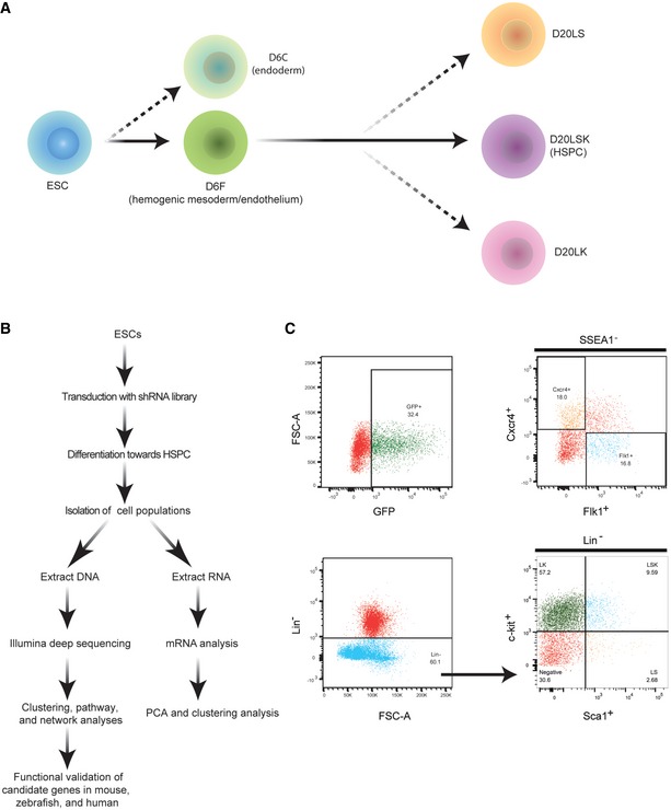
Figure 1. General outline of the experimental strategy.

- Developmental stages of ESC differentiation to HSPCs, showing the populations isolated on days 6 and 20.
- Schematic overview of the experimental strategy.
- FACS plots from a representative experiment showing gates for purification of the cell populations. Upper left: GFP+ cells in the population of ESCs infected with pGIPZ‐shRNA library lentiviruses. Upper right: Isolation of mesodermal (SSEA1−Flk1+Cxcr4−) and endodermal (SSEA1−Flkl−Cxcr4+) cells. Lower panels: Isolation of D20LS (Lin−Sca1+c‐Kit−), D20LK (Lin−Sca1−c‐Kit+), and D20LSK (Lin−Sca1+c‐Kit+) cells.
