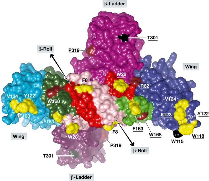
Figure 1. Three‐dimensional structure of the ZIKV NS1 dimer.

View onto the presumable membrane‐interacting (inner) surface involving the wing and the β‐roll domains of each protomer. The β‐roll domains are pink (protomer A) and red (protomer B), the wing domains are light blue (A) and dark blue (B), the connector subdomains are dark green (A) and light green (B), and the β‐ladder domains (outer surface) are faded dark purple (A) and purple (B). Residues believed to be in contact with the ER membrane are yellow; those proposed to be involved in RNA replication and virion assembly (Scaturro et al, 2015) are brown and black, respectively. Residue numbers from protomer B are underlined. Figure prepared by Dr. Jian Lei using UCSF Chimera and PDB entry 5K6K (Brown et al, 2016).
