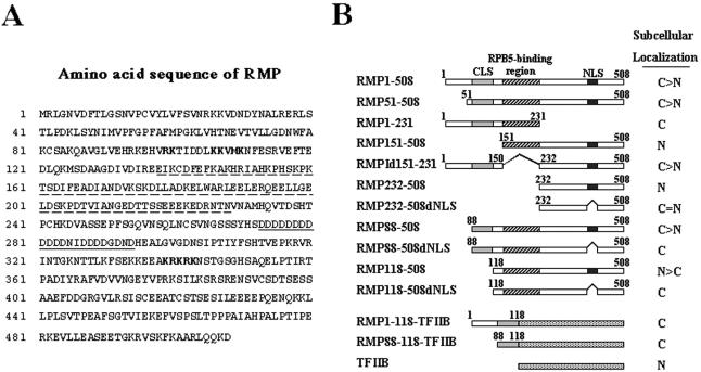
FIG. 1.
Subcellular localization of RMP. (A) Amino acid sequence of RMP. The RPB5-binding region is underlined with a dashed line, the Asp-rich region is underlined, and two different putative NLSs are shown in boldface. (B) Schematic presentation of different truncated mutants of RMP in GFP-fused form. The subcellular localization of the fusion proteins elucidated by confocal microscopy is shown at the right (C, exclusively cytoplasmic; N, exclusively nuclear; >, mostly or strongly; =, equally). The numbers indicate the number of amino acids in a given construct.

