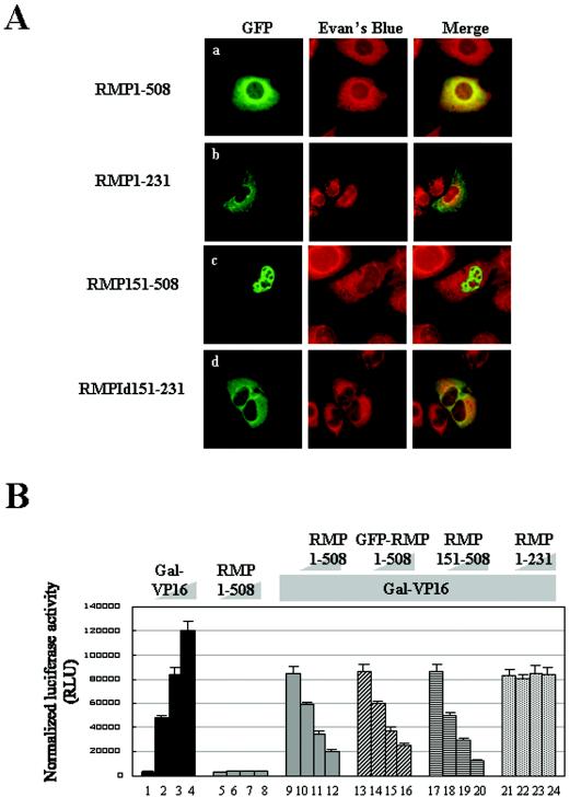
FIG. 2.
Subcellular localizations and corepressor activities of RMP and its truncated mutants. (A) HLE cells transfected with the indicated GFP-RMP constructs were stained with Evans Blue to visualize the cell structure and were observed by confocal microscopy. The expression of GFP-RMP proteins was detected by green fluorescence. (B) Corepressor activity was addressed by a dual luciferase assay as described in Materials and Methods. Various amounts of Gal-VP16 and RMP constructs were transfected together with 200 ng of reporter luciferase and 20 ng of control luciferase constructs, respectively, for each well. The cotransfection mixture contained the following constructs: bars 1 to 4, 0, 0.2, 0.4, and 0.8 ng of Gal-VP16, respectively; bars 5 to 8, 0, 0.1, 0.5, and 1.0 μg of RMP1-508, respectively; bars 9 to 12 0, 0.1, 0.5, and 1.0 μg of RMP1-508, respectively, plus 0.4 ng of Gal-VP16; bars 13 to 16, 0, 0.5, 1.0, and 2.0 μg of GFP-RMP1-508, respectively, plus 0.4 ng of Gal-VP16; bars 17 to 20, 0, 0.1, 0.5, and 1.0 μg of RMP151-508, respectively, plus 0.4 ng of Gal-VP16; and bars 21 to 24, 0, 0.1, 0.5, and 1.0 μg of RMP1-231, respectively, plus 0.4 ng of Gal-VP16. The error bars indicate standard deviations.

