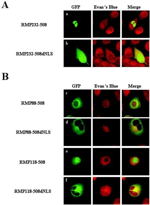
FIG. 3.
Mapping of CLS in an N-terminal region and the functional role of the NLS in the C terminus of RMP. HLE cells transfected with the indicated GFP-RMP constructs were fixed and counterstained with Evans Blue to visualize the cell structure and were observed by confocal microscopy. The expression of GFP-RMP proteins was detected by green fluorescence.

