
Figure EV2. Small clones remaining at 24 weeks harbour cells in the basal layer.
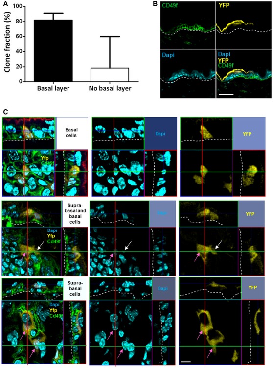
- Histogram chart showing the relative frequency of small clones observed at 24 weeks with or without cells in the basal layer. Clones without cells in the basal layer are considered as dying clones entering fully terminal differentiation. About 80% of small clones had cells in the basal layer of the epidermis (black bar), while 20% did not (white bar). n = 41 clones were counted from 4 mice. Data are represented as median with interquartile range.
- Confocal analysis of alpha‐6 integrin staining to identify basal epidermal cells. An optical section of a Z‐stack transversal acquisition demonstrates the presence of YFP+ cells above the basal layer stained with alpha‐6 integrin. This YFP+ cell is therefore suprabasal (scale bar represents 50 μm).
- Photomicrographs represent 2D optical section from a Z‐stack acquisition in of whole‐mounted skin displaying representative basal (white arrows) and suprabasal (pink arrows) clones at D3 of tamoxifen injection (scale bar represents 20 μm).