
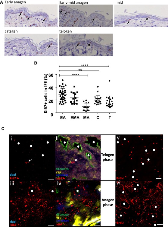
Figure 4. Proliferation in the IFE is hair cycle dependent and occurs mainly during early anagen phase.
-
A, BKi67 immunohistochemistry of back skin sections at different phases of the hair follicle cycle determined using a bioluminescent reporter mice of the canonical Wnt signalling pathway (A; scale bar represents 100 μm) and corresponding scatter plot quantification (B). Analysis focused on the basal layer of the IFE. Each dot represents one image. Proliferation in the IFE is significantly increased during the anagen phase except in mid‐anagen. All data are represented as mean ± s.e.m. Unpaired t‐test, ****P < 0.0001, **P = 0.0020. EA n = 32, EMA n = 13, MA n = 15, C n = 21, T n = 19 from 6 mice. Black arrows: Ki67‐positive cells.
-
CPhotomicrographs represent 2D optical sections from Z‐stack acquisition of whole‐mounted back skin stained in anagen and telogen with anti‐Ki67 antibody or upon BrdU incorporation. Ki67+ cells are labelled with Alexa 647 (red). The density of Ki67+ cells increased in the anagen phase (iii and iv) compared to telogen (i and ii). White arrows: Ki67+ cells. (scale bar represents 20 μm). Similarly, short‐term BrdU pulse (2 h) resulted in significant staining of anagen epidermal areas (vi, red nuclear stain) reinforcing hair follicle openings as compared to telogen areas.