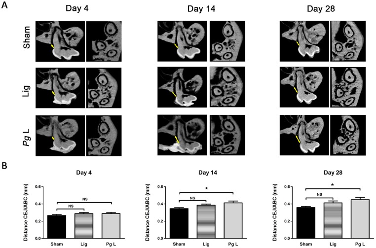
Fig 4. Time-course of alveolar bone loss in the ligature-induced murine model of experimental periodontitis.
CD1 Swiss mice (n = 90) were subjected to experimental periodontitis for 4, 14 and 28 days. At each time point, animals were sacrificed and maxillary samples were harvested. A. After 4, 14 and 28 days, μCT analysis was performed. Longitudinal sections through the middle of the palatal root of the first maxillary molar (left images) and transversal sections from the apices of the three roots of the first maxillary molar to the summit of the alveolar bone crest (right images) are presented for each time points. B. Alveolar bone loss was assessed using 2D μCT. At each time point, data of ligatured groups (Lig and Pg L) were compared to their respective Sham groups. Data are shown as means ± SEM. * p<0.05.

