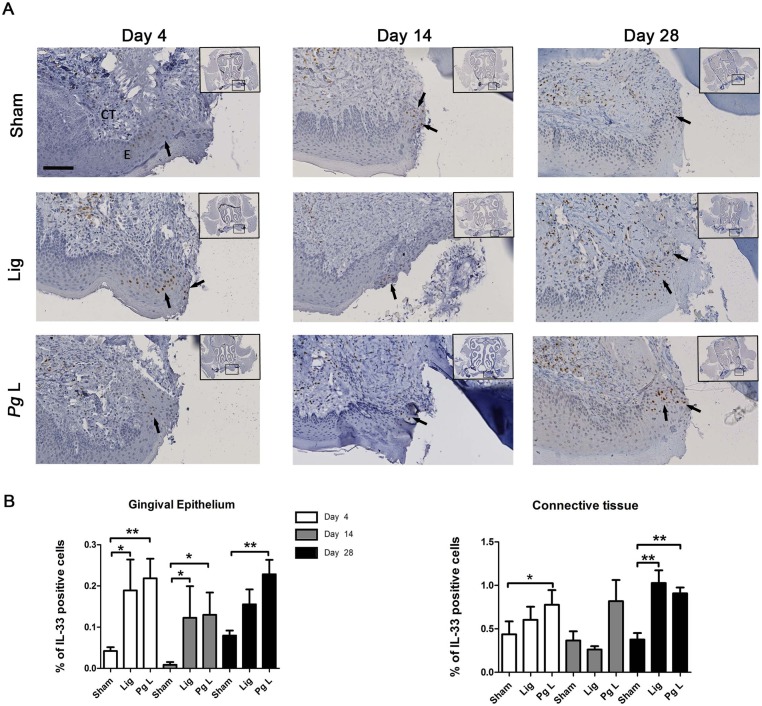
Fig 5. Time-course of IL-33 expression in the ligature-induced murine model of experimental periodontitis.
A. IL-33 expression was assessed by IHC and sections were counterstained with Harris Hematoxylin staining (arrows). B. The percentage of IL-33 was quantified in gingival epithelium and in connective tissue using Fiji software and defined as a percentage of DAB positive staining area per region of interest. At each time point, data of ligatured groups (Lig and Pg L) were compared to their respective Sham groups. EP: Epithelium, CT: Connective tissue. Data are shown as means ± SEM. * p<0.05; **p<0.01. Scale bar = 100μm.

