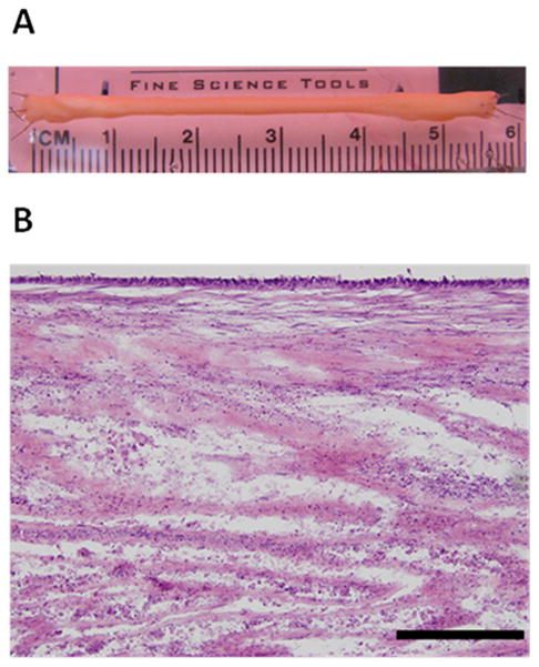
Figure 1.

In Vitro BLB (A) Representative image of BLB. Each ACL BLB construct was fabricated with 2-5 smaller BLBs made of a 30 mm ligament portion flanked by two 15 mm bone ends. These constructs were placed side-by-side and allowed to fuse in vitro before implantation. (B) Representative longitudinal H&E section of a BLB in vitro prior to implantation. Scale bar indicates 200 μm.
