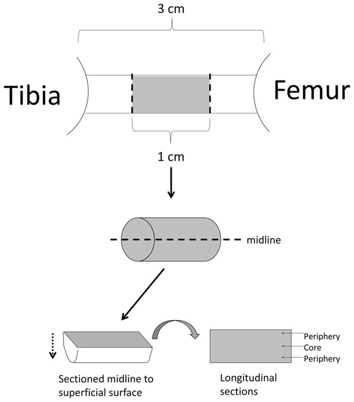
Figure 2.
Sectioning Procedure for Histological Analysis: Immunohistological quantification was achieved by first taking the middle 1 cm of the tissue spanning the intra-articular space and splitting this tissue longitudinally at the midline into two samples to expose the entire mid portion of the graft, including tissue from the periphery and core. The samples were sectioned at 12 μm starting from the midline and moving towards the superficial surface, taking sections for 6 separate stains.

