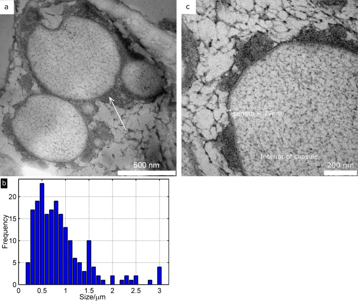
Fig 2. Morphology of capsules.
(a) TEM micrographs of the synthesized capsules showing the capsule structure. The arrow points to a darker area, most likely diisocyanate debris from capsule synthesis. (b) Histogram showing the distribution in capsule size. (c) A close-up of a synthesized capsule, showing the cell wall (marked) and the structure of the interior of the capsule.

