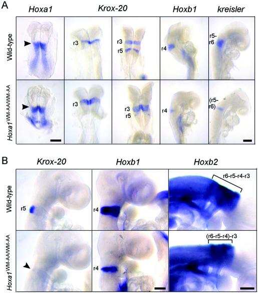
FIG. 2.
Analysis of hindbrain patterning by whole-mount in situ hybridization. (A) Wild-type (top) and mutant (bottom) embryos between 7.5 and 8.75 dpc hybridized with a probe for Hoxa1, Krox-20, Hoxb1, or kreisler. The arrowheads indicate the anterior limits of expression of Hoxa1, corresponding to the presumptive r3-r4 boundary. In the mutant, the r3 expression domain of Krox-20 is enlarged, while its r5 expression domain is drastically reduced. Moreover, the r4 expression domain of Hoxb1 is reduced, and the kreisler r5-r6 domain of expression is only one rhombomere long. (B) Wild-type (top) and mutant (bottom) 9.5-dpc embryos hybridized with a probe for Krox-20, Hoxb1, or Hoxb2. In the mutant, the r5 expression domain of Krox-20 is almost absent (arrowhead), the r4 expression domain of Hoxb1 is clearly reduced, and the highly stained portion of the Hoxb2 expression domain, corresponding to r3-r6, is shortened (bracket). Scale bars, 200 μm.

