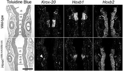
FIG. 3.
Analysis of hindbrain patterning by in situ hybridization on serial coronal sections. Expression of Krox-20, Hoxb1, and Hoxb2 on serial coronal sections of 9.5-dpc wild-type (top) and mutant (bottom) embryos. Bright-field views after toluidine blue staining are presented on the left, and dark-field views after in situ hybridization are shown on the right. The arrowhead points to the Krox-20 r5 expression domain, drastically reduced in the mutant. ov, otic vesicle. Scale bar, 200 μm.

