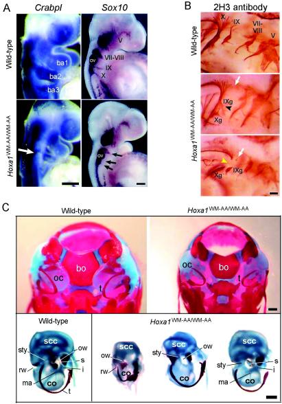
FIG. 4.
Analysis of neural crest derivatives and skeletons. (A) Whole-mount in situ hybridization on wild-type (top) and homozygous mutant (bottom) embryos at 9.25 and 9.5 dpc. The embryos were hybridized with a CrabpI or a Sox10 probe. The open arrow indicates the reduction of the ncc population expressing CrabpI and migrating into the second branchial arch (ba2). The solid arrows indicate the reduction of neurogenic ncc at the level of nerve VII-VIII and the absence of Sox10 expression at the level of nerves IX and X. ba, branchial arch; ov, otic vesicle. (B) Immunostaining of cranial nerves of a 10.5-dpc wild-type embryo (top) and two Hoxa1WM-AA/WW-AA mutant embryos (bottom) (caudal is on the left). In the mutants, the open arrows indicate the absence of nerve IX dorsal roots, the solid arrowhead indicates the fusion of the inferior ganglion of nerve IX (IXg) with the inferior ganglion of nerve X (Xg), and the yellow arrowhead points to the lack of the interganglionic portion of nerve X. (C) (Top) Dorsal view of the skull of wild-type (left) and Hoxa1WM-AA/WW-AA mutant (right) newborns. In the mutant, the shape of the basioccipital bone (bo) is altered, the otic capsules (oc) are reduced, and the tympanic ring (t) is displaced. (Bottom) Dissected otic capsules and tympanic rings from a wild-type skull (left) and three Hoxa1WM-AA/WW-AA mutant skulls (right). co, cochlea; i, incus; ma, malleus; ow, oval window; rw, round window; s, stapes; scc, semicircular canals; sty, styloid process; t, tympanic ring. Scale bars, 200 (A and B) and 500 (C) μm.

