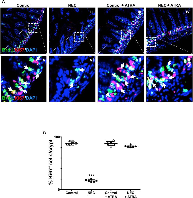
Fig. 3. Enteral administration of retinoic acid preserves the proliferative capacity of crypt-based intestinal stem cells.
A, the proliferative capacity of crypt-based ISCs was assessed by BrdU label incorporation and Ki67 staining of terminal ileum sections obtained from control mice that did not (i and v) or did (iii and vii) receive ATRA (50 μg/mouse orally per day) and mice submitted to experimental NEC in the absence (ii and vi) or presence (iv and viii) of ATRA. The outlined areas in i – iv are shown at a higher magnification in v – viii. Proliferating cells are indicated by arrows and demonstrate a conserved stable pool of dividing cells within the crypts. B, Ki67 staining was quantified as described in the methods section and is expressed as a percentage of the total number of cells identified within the crypts per high-magnification field. Scale bar = 50μm. Data depicted is presented as mean ± SD and represents 3 independent experiments with at least 10 mice per group. ***P ≤ 0.0001 determined by ANOVA followed by Tukey's multiple comparisons.

