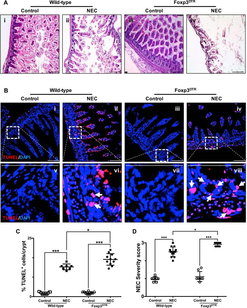
Fig. 6. Depletion of regulatory T cells exacerbates the development of experimental necrotizing enterocolitis.
A, H&E sections of the terminal ileum obtained from wild-type C57Bl/6 and Foxp3+DTR mice submitted to experimental NEC (ii and iv, respectively) or breast-fed controls (i and iii, respectively). B, confocal images of TUNEL stained sections of the terminal ileum obtained from wild-type C57Bl/6 and Foxp3+DTR mice submitted to experimental NEC (ii, vi and iv, viii respectively) or breast-fed controls (i, v and iii, vii respectively). The outlined areas in i – iv are shown at a higher magnification in v – viii. Apoptotic cells within the crypts are identified by arrows and demonstrate the significant effect that depletion of Foxp3+ T cells has on the induction and severity of NEC, exacerbating the damage to the crypt-based ISC population. TUNEL staining intensity (C) was quantified as described in the methods section and is expressed as a percentage of the total number of cells identified within the crypts per high-magnification field. D, injury severity score as determined by a pathologist blinded to the experimental conditions, demonstrates a significant increase in the incidence and severity of experimental NEC among Foxp3+DTR mice. Scale bar = 50μm. Data depicted is presented as mean ± SD and represents 3 independent experiments with at least 10 mice per group. *P ≤ 0.05, ***P ≤ 0.0001 determined by ANOVA followed by Tukey's multiple comparisons.

