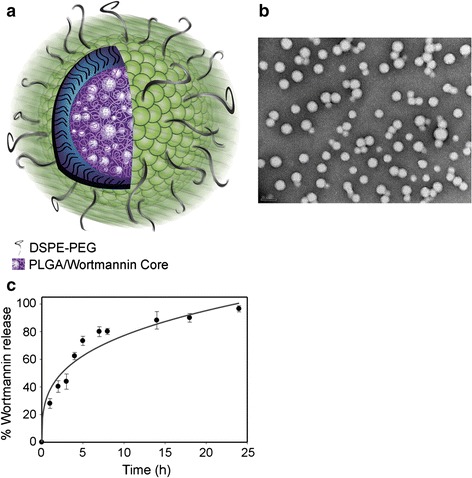
Fig. 1.

Characterization of NP Wtmn. a Cartoon of NP Wtmn depicting a PLGA core containing Wtmn surrounded by a lipid monolayer (green head groups) and a PEG shell. b TEM image of NP Wtmn. c Release profile of NP Wtmn in PBS at 37 °C. Error bars correspond to SD of three separate sample preparations with duplicate samples per data point (Karve et al. 2012)
