Figure 4.
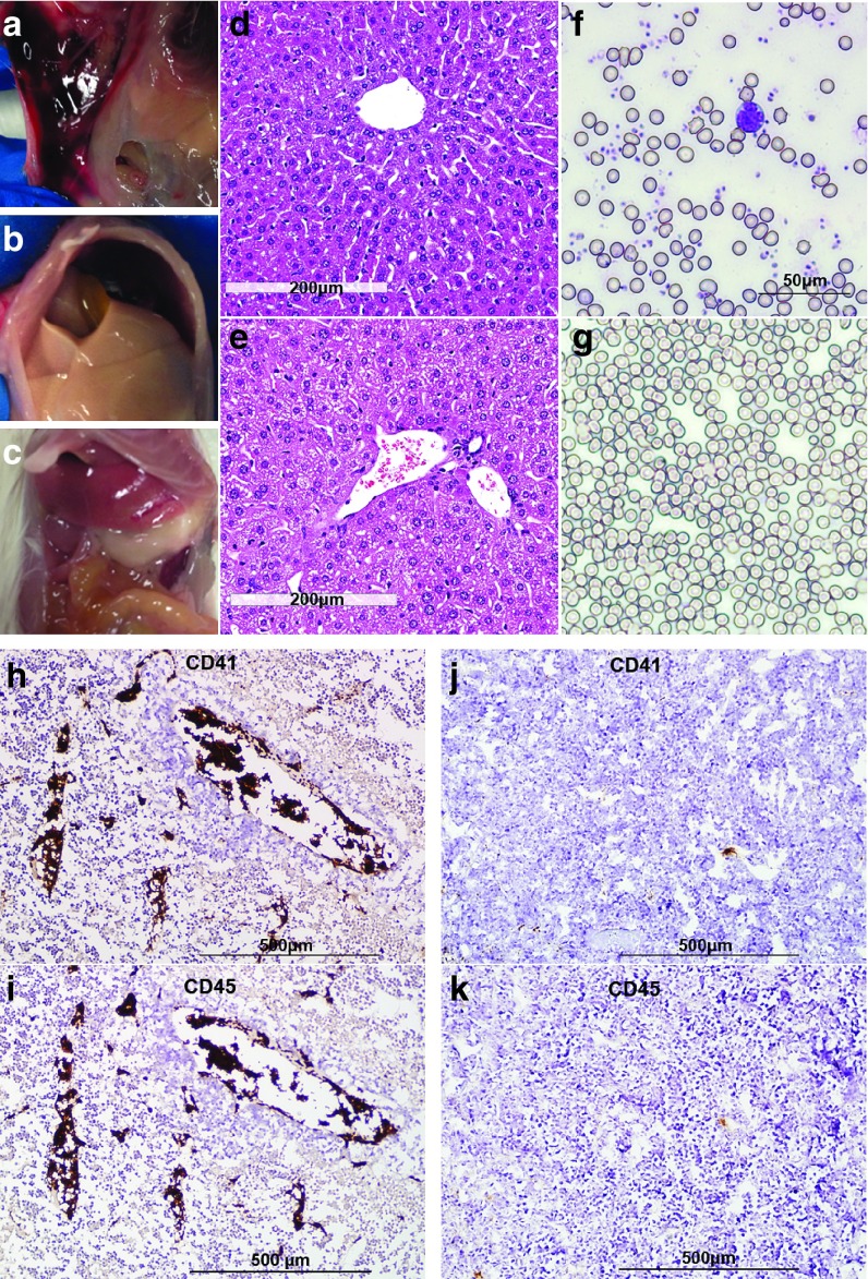
Histological analysis of blood and tissues from vesicular stomatitis viruses (VSV)-treated MPC-11 bearing mice suggest intravascular coagulation and consumptive coagulopathy. Photographs of VSV treated mice taken at necropsy showed cases of (a) abdominal wall bleeding, and (b) pale ischemic liver. (c) A normal liver is shown for comparison. H&E stained liver sections from (d) saline-treated or (e) VSV-treated MPC-11 mice showing fibrin deposits and trapped erythrocytes in vessels. (f) Erythrocytes and platelets were seen in May-Grünwald-Giemsa stained blood smears from saline-treated mice. (g) Platelets were noticeably reduced in blood smears of VSV-treated mice at day 4 post-treatment. Immunohistochemical staining for platelets (CD41) and lymphocytes (CD45) in tumors harvested from (h, i) VSV-treated and (j, k) saline-treated mice. Aggregates of platelets and lymphocytes, as a result of intravascular coagulation, can be seen in the blood vessels of VSV-treated animals at day 4.

