Figure 2.
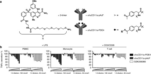
Characterization of the anti-inflammatory effect of αhuCD11a-PDE4 ADC in vitro. (a) 3-linker was synthesized based on compound 3 for antibody conjugation. This PDE4 inhibitor linker derivative was then conjugated to αhuCD11a-pAcF to generate αhuCD11a-PDE4 ADC. (b) Freshly isolated human PBMCs, monocytes and T cells were treated with various concentrations of αhuCD11a-PDE4, αhuCD11a-pAcF, and GSK256066. The highest concentration on right side for αhuCD11a-PDE4 and αhuCD11a-pAcF was 100 nmol/l, and for GSK256066 was 30 nmol/l. All treatments from right to left are in 1:3 serial dilution. After 7-hour incubation, PBMCs and monocytes were treated with 100 ng/ml LPS and T cells were treated with CD3/CD28 Dynabeads in 1:1 ratio. After 20-hour stimulation, TNFα levels in the cell culture supernatants were assessed by an HTRF assay. TNFα levels were normalized to LPS or CD3/CD28 treatment alone. Results are presented as means ± SD.

