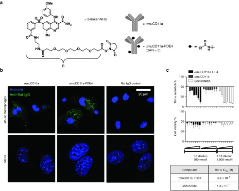
Figure 3.
Characterization of the anti-inflammatory effect of αmuCD11a-PDE4 ADC in vitro. (a) PDE4 inhibitor linker derivative (3-linker-NHS) basing on compound 3 was conjugated to αmuCD11a to generate αmuCD11a-PDE4 ADC. (b) Mouse primary peritoneal cells and mouse embryonic fibroblasts (MEFs) were incubated with αmuCD11a-PDE4, αmuCD11a or Rat IgG control for 7 hours before being fixed and stained with an anti-rat IgG secondary antibody conjugated with Alexa 488 (green). The nuclei was stained with Hoescht (blue). The cells were imaged with confocal microscopy. (c) Mouse primary peritoneal cells were treated with various concentrations of αmuCD11a-PDE4, αmuCD11a, or GSK256066. The highest concentration on the right side for αmuCD11a-PDE4 and αmuCD11a was 900 nmol/l with 1:3 serial dilution from right to left, and for GSK256066 was 1,000 nmol/l with 1:10 serial dilution from right to left. TNFα IC50 values for αmuCD11a-PDE4 and GSK256066 in this assay were calculated and shown in a table. Results are presented as means ± SD.

