Figure 4.
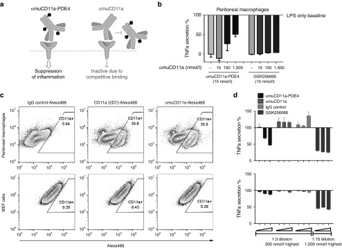
Anti-inflammatory effect of αmuCD11a-PDE4 ADC is mediated through CD11a receptor. (a) A schematic graph showing that αmuCD11a-PDE4 binds CD11a receptor and exerts its anti-inflammatory effect, while occupancy of CD11a receptor by αmuCD11a blocks αmuCD11a-PDE4 from accessing to CD11a receptor, thus abrogating αmuCD11a-PDE4's anti-inflammatory effect. (b) Mouse peritoneal cells were treated with different concentrations of αmuCD11a as indicated for 30 minutes before αmuCD11a-PDE4 or GSK256066 was added. After 7-hour incubation, cells were stimulated with 100 ng/ml LPS for 20 hours. The TNFα content in the supernatant was assessed by an HTRF assay. The results are normalized to LPS treatment alone and are presented as means ± SD. (c) Peritoneal macrophages or MEFs were stained with IgG control, CD11a (2D7) or αmuCD11a antibodies conjugated with Alexa 488 and were analyzed by flow cytometry. (d) Mouse primary peritoneal cells (upper panel) or MEFs (lower panel) were treated with various concentrations of αmuCD11a-PDE4, αmuCD11a, rat IgG control or GSK256066. The highest concentration from right side for αmuCD11a-PDE4, αmuCD11a and IgG control was 300 nmol/l with 1:3 serial dilution from right to left, and for GSK256066 was 1,000 nmol/l with 1:10 serial dilution from right to left. The results are normalized to LPS treatment alone, and are presented as means ± SD.

