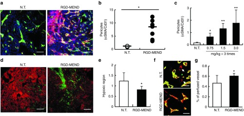
Figure 2.
Vascular maturation by siRNA against VEGFR2 encapsulated in the RGD-MEND. (a) Representative image of the increase in pericyte coverage by the RGD-MEND. Tumor tissues were cryo-sectioned after the continuous inhibition of VEGFR2. The sections were stained with Hoechst33342 (blue, nucleus), FITC-isolectin (green, vessels) and cy3-αSMA (red, pericytes). Scale bars: 100 μm. (b) Quantitative data of a. Pixels were counted in nine images from three independent mice, and the red pixels (pericytes) were then normalized to green pixels (vessels). (c) Dose-dependency for the increase in pericyte coverage. Pericytes were counted when the dosages of si-VR2 varied from 0.75 mg/kg to 3.0 mg/kg (each groups were of three mice). (d) Decrease in hypoxic area in RGD-MEND-treated mice. Tumor tissues were collected 90 minutes after the injection of the hypoxia-probe pimonidazole. Green and red pixels vessels and the indicated hypoxic regions, respectively. Scale bars: 100 μm. (e) Red dots indicating hypoxic regions were counted and normalized to nucleus areas. Data were obtained from nine images from three independent mice. (f) Recovery of blood flow by the RGD-MEND. FITC-isolectin B4 was systemically injected before sacrifice, and the collected tumor tissues were then immersed in Alexa647-isolectin B4. Arrows show the vasculature without blood flow. (g) Quantitative data of perfused vessels. Population of the vasculature with blood flows (shown as yellow) against all of the vasculature (shown as yellow and red) were counted. FITC, fluorescein isothiocyanate; RGD-MEND, RGD-modified liposomal siRNA; VEGFR2, VEGF receptor 2.

