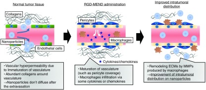
Figure 5.
Conceptual illustration of the improvement of intratumoral distribution of nanoparticles. In untreated tumor tissue, the tumor vasculature is immature. Specifically the vasculature lacks pericyte coverage and basement membrane and fenestrae (intracellular pore) and a loose junction (intercellular gap) exists. For these reasons, nanoparticles can pass through the vascular wall, a process that is called the EPR effect. However, the presence of abundant collagen molecules restrict the intratumoral diffusion of the nanoparticles. This study revealed that the inhibition of VEGFR2 on tumor endothelial cells (TECs) by the RGD-MEND leads to the infiltration of macrophages. The macrophages then produce matrix metalloproteinases (MMPs) that catalyze the degradation of the extracellular matrices (ECMs). After the remodeling of the ECMs, nanoparticles are able to penetrate more deeply into the tumor tissue. EPR, enhanced permeability and retention; RGD-MEND, RGD-modified liposomal siRNA; VEGFR2, VEGF receptor 2.

