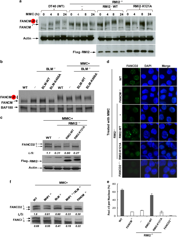
Figure 5.
RMI2-mediated association between FANCM and BLM complex is required for FANCM hyperphosphorylation, FANCD2 monoubiquitination and foci formation. (a) Immunoblotting shows that FANCM hyperphosphorylation and FANCD2 monoubiquitination are concomitantly reduced in MMC-treated RMI2−/− cells, or RMI2−/− cells expressing RMI2-121A point mutant. Wild-type, or RMI2−/− cells, or RMI2−/− cells complemented by either wild-type RMI2 or 121A point mutant [43], were treated with MMC (50 ng ml−1) for increasing lengths of time, as indicated above the images. They were then collected for western analyses. The monoubiquitinated and non-ubiquitinated FANCD2 was indicated as FANCD2-L and S (long and short), whereas hyperphosphorylated FANCM was marked as FANCM-P and was blotted from 6% gel with extended electrophoresis. The mobility of FANCM is decreased in response to MMC due to hyperphosphorylation [19]. (b) Immunoblotting shows that the level of hyperphosphorylated FANCM is modestly reduced in BLM mutant DT40 cells treated with MMC (50 ng ml−1) for 18 h. Immunoblotting of BAF180 (a subunit of PBAF chromatin remodeling complex) was included as a loading control. (c) Immunoblotting shows the level of monoubiquitinated FANCD2 is reduced in RMI2−/− cells, or in the same cells expressing RMI2-K121A mutant. The cells were all treated with MMC (50 ng ml−1) for 18 h. The ratio between the monoubiquitinated and ubiquitinated FANCD2 (L/S) was shown. (d) Representative images and (e) their quantification show that MMC-induced FANCD2 focus formation is reduced in MMC-treated RMI2−/− cells, or RMI2−/− cells expressing RMI2-121A point mutant. Various DT40 cell lines were treated with MMC (60 ng ml−1) for 18 h before they were collected for the analyses of FANCD2 foci. The cells include wild-type (WT), RMI2−/−, RMI2−/− cells complemented with RMI2 wild-type (WT) or RMI2-K121A mutant, FANCM−/− and FANCD2−/− cells. The latter two cells were included as controls. The graph in e shows the mean and standard deviations (s.d.) of the percentage of cells containing ⩾5 FANCD2 foci, as assayed in d. The P-values between different cell lines are shown. (f) Immunoblotting shows the levels of monoubiquitinated FANCD2 and FANCI in various DT40 cell lines as indicated. The cells were all treated with MMC (50 ng ml−1) for 18 h. The ratios between the monoubiquitinated and ubiquitinated FANCD2 or FANCI (L/S) were shown. MMC, mitomycin C.

