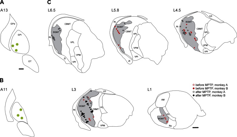
Fig. 2.
Stimulation sites in the internal globus pallidus (GPi, A and B) and microelectrode recording sites in the BGMT (C). The drawings in A and B are in coronal planes, showing the stimulation electrode tip sites (green circles) in monkeys A and B, respectively. The drawing of recording sites in C is based on histological reconstructions of parasagittal Nissl-, calbindin-, and MAP2-stained slides. Data from both monkeys are combined. The nuclear boundaries in A and B are based on the nuclear outlines in the atlas by Paxinos et al. (Paxinos et al. 2000), and those in C are drawn using the macaque brain atlas by Lanciego et al. (Lanciego and Vazquez 2012). In C, neurons recorded before 1-methyl-4-phenyl-1,2,3,6-tetrahydropyridine (MPTP) treatment are shown as red symbols, neurons recorded after MPTP treatment are shown in black. Cells that were very closely spaced are shown next to each other. Open symbols represent recordings in monkey A, filled symbols are from monkey B. AM, anterior medial nucleus of the thalamus; CBMT, cerebellar receiving territory of the motor thalamus; GPe, external pallidal segment; LD, laterodorsal nucleus of the thalamus; OT, optic tract; PC, paracentral nucleus of the thalamus; Re, reuniens nucleus of the thalamus; Rt, reticular nucleus of the thalamus; VPI, ventral posterior inferior nucleus of the thalamus; VPL, ventral posterior lateral nucleus of the thalamus; VPM, ventral posterior medial nucleus of the thalamus; ZI, zona incerta. Scale bars, 1 mm. The scale bar in A also applies to B.

